The right and left medial pterygoid plates form the posterior lateral walls of the nasal cavity. It may be absent or asymmetrical in some people.
Medial Pterygoid Muscle Attachments Actions Innervation
A central consisting of the body and small wings and two lateral each comprising a great wing.
. The smaller superficial head originates from the. The risorius muscle is a muscle of facial expression. The medial pterygoid muscle is a thick muscle that is located from the back of the molars to just under eye level it is located behind the orbits.
It is located inferiorly to the lateral pterygoid. About the fourth month a center appears for each lingula and speedily joins the rest of the bone. These are the medial pterygoid plate and lateral pterygoid plate pterygoid wing-shaped.
The risorius muscle arises in the fascia over the parotid gland. The larger deep head arises from the medial surface of the lateral pterygoid plate of the sphenoid bone and the adjacent pyramidal process of palatine bone. The outer surface of the muscle lies against the inner surface of mandible from which it is separated by the lateral pterygoid muscle sphenomandibular ligament maxillary artery mandibular nerve and its.
It retracts the angle of the mouth during smiling. It is composed of two heads that have two sets of origins. The medial joins the lateral pterygoid plate about the sixth month.
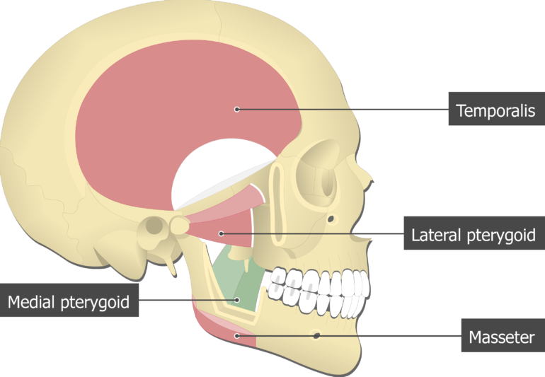
It arises from the fascia over the parotid gland and inserts into the angle of the mouth. The deep head originates from the medial aspect of the lateral pterygoid plate of the. Medial pterygoid muscle is located in the infratemporal fossa lying deep to masseter and temporalis muscles and medial to lateral pterygoid muscle.
The somewhat larger lateral pterygoid plates serve as attachment sites for chewing muscles that fill the infratemporal space and act on the mandible. The superficial head originates from the maxillary tuberosity and the pyramidal process of palatine bone. The presphenoid is united to the postsphenoid about the eighth month and at birth the sphenoid is in three pieces Fig.
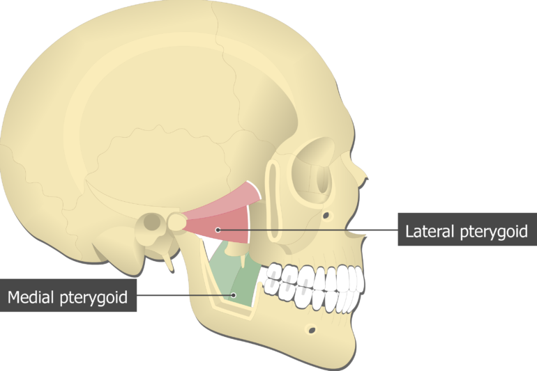
The medial pterygoid muscle has a quadrangular shape with two heads. The medial pterygoid muscle is a quadrangular muscle situated in the infratemporal fossa. It has many functions including closing the jaw moving the jaw back to the middle if excursion side-to-side movement has occurred and aiding in protrusion of the mandible which is when the jaw moves forward.
It is supplied by the facial nerve CN VII.
Medial Pterygoid Muscle Attachments Actions Innervation

Medial Pterygoid Muscle Wikipedia
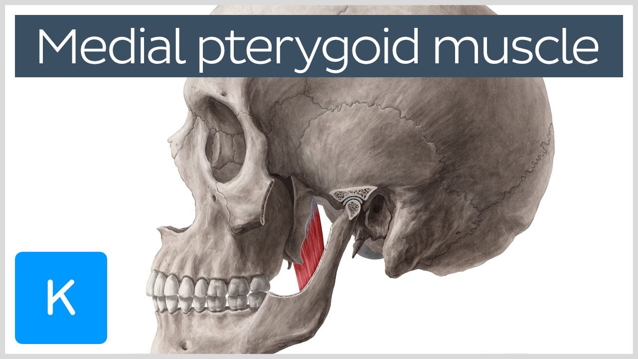
Medial Pterygoid Muscle Origin Insertion Function Nerve Supply Anatomy Kenhub Youtube

Figure Medial Pterygoid Muscle Image Courtesy O Chaigasame Statpearls Ncbi Bookshelf
![]()
Medial Pterygoid Origin Insertion Action Innervation Kenhub
![]()
Medial And Lateral Pterygoid Muscle Anatomy And Function Kenhub

Medial Pterygoid Muscle Origin And Insertion Google Search Human Anatomy And Physiology Muscle Anatomy Muscular System
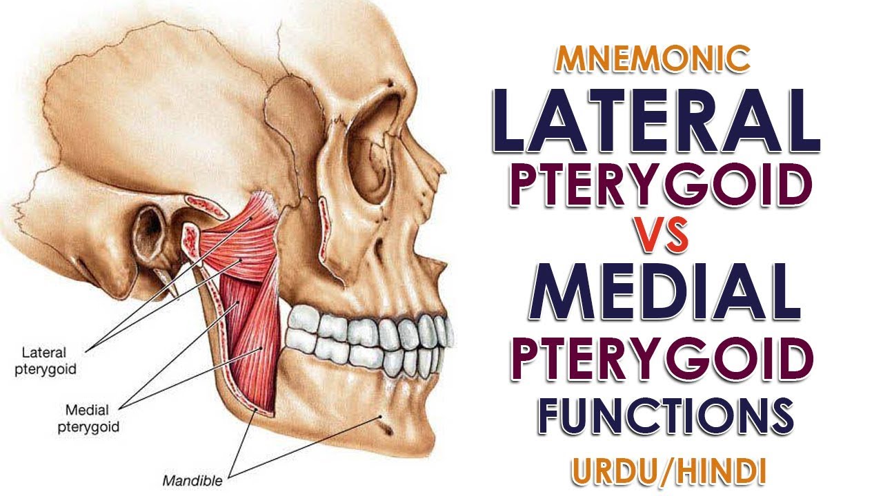
Mnemonic Lateral Pterygoid Vs Medial Pterygoid Function Urdu Hindi Youtube
0 comments
Post a Comment